Everything About Fungus 1

Importance of fungi
Humans have been indirectly aware of fungi since the first loaf of leavened bread was baked and the first tub of grape must was turned into wine. Ancient peoples were familiar with the ravages of fungi in agriculture but attributed these diseases to the wrath of the gods. The Romans designated a particular deity, Robigus, as the god of rust and, in an effort to appease him, organized an annual festival, the Robigalia, in his honour.
Fungi are everywhere in very large numbers—in the soil and the air, in lakes, rivers, and seas, on and within plants and animals, in food and clothing, and in the human body. Together with bacteria, fungi are responsible for breaking down organic matter and releasing carbon, oxygen, nitrogen, and phosphorus into the soil and the atmosphere. Fungi are essential to many household and industrial processes, notably the making of bread, wine, beer, and certain cheeses. Fungi are also used as food; for example, some mushrooms, morels, and truffles are epicurean delicacies, and mycoproteins (fungal proteins), derived from the mycelia of certain species of fungi, are used to make foods that are high in protein.
Studies of fungi have greatly contributed to the accumulation of fundamental knowledge in biology. For example, studies of ordinary baker’s or brewer’s yeast (Saccharomyces cerevisiae) led to discoveries of basic cellular biochemistry and metabolism. Some of these pioneering discoveries were made at the end of the 19th century and continued during the first half of the 20th century. From 1920 through the 1940s, geneticists and biochemists who studied mutants of the red bread mold, Neurospora, established the one-gene–one-enzyme theory, thus contributing to the foundation of modern genetics. Fungi continue to be useful for studying cell and molecular biology, genetic engineering, and other basic disciplines of biology.
The medical relevance of fungi was discovered in 1928, when Scottish bacteriologist Alexander Fleming noticed the green mold Penicillium notatum growing in a culture dish of Staphylococcus bacteria. Around the spot of mold was a clear ring in which no bacteria grew. Fleming successfully isolated the substance from the mold that inhibited the growth of bacteria. In 1929 he published a scientific report announcing the discovery of penicillin, the first of a series of antibiotics—many of them derived from fungi—that have revolutionized medical practice.
Another medically important fungus is Claviceps purpurea, which is commonly called ergot and causes a plant disease of the same name. The disease is characterized by a growth that develops on grasses, especially on rye. Ergot is a source of several chemicals used in drugs that induce labour in pregnant women and that control hemorrhage after birth. Ergot is also the source of lysergic acid, the active principle of the psychedelic drug lysergic acid diethylamide (LSD). Other species of fungi contain chemicals that are extracted and used to produce drugs known as statins, which control cholesterol levels and ward off coronary heart disease. Fungi are also used in the production of a number of organic acids, enzymes, and vitamins.
Form and function of fungi
Size range
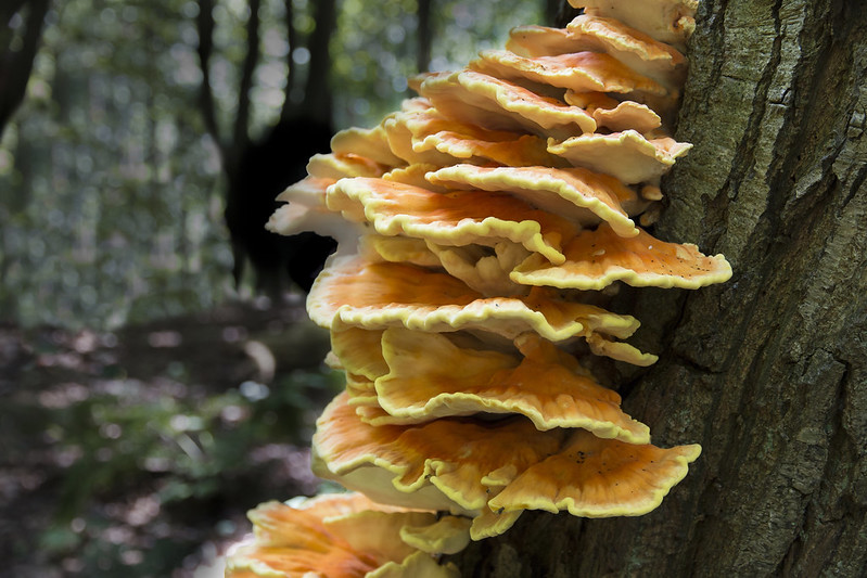
The mushrooms, because of their size, are easily seen in fields and forests and consequently were the only fungi known before the invention of the microscope in the 17th century. The microscope made it possible to recognize and identify the great variety of fungal species living on dead or live organic matter. The part of a fungus that is generally visible is the fruiting body, or sporophore. Sporophores vary greatly in size, shape, colour, and longevity. Some are microscopic and completely invisible to the unaided eye; others are no larger than a pin head; still others are gigantic structures. Among the largest sporophores are those of mushrooms, bracket fungi, and puffballs. Some mushrooms reach a diameter of 20 to 25 cm (8 to 10 inches) and a height of 25 to 30 cm (10 to 12 inches). Bracket, or shelf, fungi can reach 40 cm (16 inches) or more in diameter. A specimen of the bracket fungus Fomitiporia ellipsoidea discovered in 2010 on Hainan Island in southern China had a fruiting body measuring 10.8 metres (35.4 feet) in length and 82–88 cm (2.7–2.9 feet) in width. It may have held some 450 million spores and weighed an estimated 400–500 kg (882–1,102 pounds), at the time making it the largest fungal fruiting body ever documented. Puffballs also can grow to impressive sizes. The largest puffballs on record measured 150 cm (5 feet) in diameter. The number of spores within such giants reaches several trillion.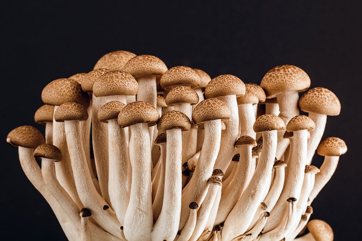
Distribution and abundance
Fungi are either terrestrial or aquatic, the latter living in freshwater or marine environments. Freshwater species are usually found in clean, cool water because they do not tolerate high degrees of salinity. However, some species are found in slightly brackish water, and a few thrive in highly polluted streams. Soil that is rich in organic matter furnishes an ideal habitat for a large number of species; only a small number of species are found in drier areas or in habitats with little or no organic matter. Fungi are found in all temperate and tropical regions of the world where there is sufficient moisture to enable them to grow. A few species of fungi live in the Arctic and Antarctic regions, although they are rare and are more often found living in symbiosis with algae in the form of lichens (see below Lichens). About 144,000 species of fungi have been identified and described, but mycologists estimate that there may be between 2.2 million and 3.8 million total species.
Basic morphology
A typical fungus consists of a mass of branched, tubular filaments enclosed by a rigid cell wall. The filaments, called hyphae (singular hypha), branch repeatedly into a complicated, radially expanding network called the mycelium, which makes up the thallus, or undifferentiated body, of the typical fungus. The mycelium grows by utilizing nutrients from the environment and, upon reaching a certain stage of maturity, forms—either directly or in special fruiting bodies—reproductive cells called spores. The spores are released and dispersed by a wide variety of passive or active mechanisms; upon reaching a suitable substrate, the spores germinate and develop hyphae that grow, branch repeatedly, and become the mycelium of the new individual. Fungal growth is mainly confined to the tips of the hyphae, and all fungal structures are therefore made up of hyphae or portions of hyphae.
Some fungi, notably the yeasts, do not form a mycelium but grow as individual cells that multiply by budding or, in certain species, by fission. In addition, the so-called cryptomycota, a primitive group of microscopic fungi, diverge significantly from the standard body plan of other fungi in that their cell walls lack the rigid polymer known as chitin. These microscopic fungi also possess a whiplike flagellum.
Structure of the thallus
In almost all fungi the hyphae that make up the thallus have cell walls. (The thalli of the true slime molds lack cell walls and, for this and other reasons, are classified as protists rather than fungi.) A hypha is a multibranched tubular cell filled with cytoplasm. The tube itself may be either continuous throughout or divided into compartments, or cells, by cross walls called septa (singular septum). In nonseptate (i.e., coenocytic) hyphae the nuclei are scattered throughout the cytoplasm. In septate hyphae each cell may contain one to many nuclei, depending on the type of fungus or the stage of hyphal development. The cells of fungi are similar in structure to those of many other organisms. The minute nucleus, readily seen only in young portions of the hypha, is surrounded by a double membrane and typically contains one nucleolus. In addition to the nucleus, various organelles—such as the endoplasmic reticulum, Golgi apparatus, ribosomes, and liposomes—are scattered throughout the cytoplasm.
Hyphae usually are either nonseptate (generally in the more primitive fungi) or incompletely septate (meaning that the septa are perforated). This permits the movement of cytoplasm (cytoplasmic streaming) from one cell to the next. In fungi with perforated septa, various molecules are able to move rapidly between hyphal cells, but the movement of larger organelles, such as mitochondria and nuclei, is prevented. In the absence of septa, both mitochondria and nuclei can be readily translocated along hyphae. In mating interactions between filamentous Basidiomycota, the nuclei of one parent often invade the hyphae of the other parent, because the septa are degraded ahead of the incoming nuclei to allow their passage through the existing hyphae. Once the incoming nuclei are established, septa are re-formed.
Variations in the structure of septa are numerous in the fungi. Some fungi have sievelike septa called pseudosepta, whereas fungi in other groups have septa with one to few pores that are small enough in size to prevent the movement of nuclei to adjacent cells. Basidiomycota have a septal structure called a dolipore septum that is composed of a pore cap surrounding a septal swelling and septal pore. This organization permits cytoplasm and small organelles to pass through but restricts the movement of nuclei to varying degrees.
The wall of the hypha is complex in both composition and structure. Its exact chemical composition varies in different fungal groups. In some funguslike organisms the wall contains considerable quantities of cellulose, a complex carbohydrate that is the chief constituent of the cell walls of plants. In most fungi, however, two other polymers—chitin and glucan (a polymer of glucose linked at the third carbon and branched at the sixth), which forms an α-glucan layer and a special β-1,3-1,6-glucan layer—form the main structural components of the wall. Among the many other chemical substances in the walls of fungi are some that may thicken or toughen the wall of tissues, thus imparting rigidity and strength. The chemical composition of the wall of a particular fungus may vary at different stages of the organism’s growth—a possible indication that the wall plays some part in determining the form of the fungus. In some fungi, carbohydrates are stored in the wall at one stage of development and are removed and utilized at a later stage. In some yeasts, fusion of sexually functioning cells is brought about by the interaction of specific chemical substances on the walls of two compatible mating types.
When the mycelium grows in or on a surface, such as in the soil, on a log, or in culture medium, it appears as a mass of loose, cottony threads. The richer the composition of the growth medium, the more profuse the threads and the more feltlike the mass. On the sugar-rich growth substances used in laboratories, the assimilative (somatic) hyphae are so interwoven as to form a thick, almost leathery colony. On the soil, inside a leaf, in the skin of animals, or in other parasitized plant or animal tissues, the hyphae are usually spread in a loose network. The mycelia of the so-called higher fungi does, however, become organized at times into compact masses of different sizes that serve various functions. Some of these masses, called sclerotia, become extremely hard and serve to carry the fungus over periods of adverse conditions of temperature and moisture. One example of a fungus that forms sclerotia is ergot (Claviceps purpurea), which causes a disease of cereal grasses. The underground sclerotia of Wolfiporia extensa, an edible pore fungus also known as tuckahoe, may reach a diameter of 20 to 25 cm (8 to 10 inches).
Various other tissues are also produced by the interweaving of the assimilative hyphae of some fungi. Stromata (singular stroma) are cushionlike tissues that bear spores in various ways. Rhizomorphs are long strands of parallel hyphae cemented together. Those of the honey mushroom (Armillaria mellea), which are black and resemble shoestrings, are intricately constructed and are differentiated to conduct water and food materials from one part of the thallus to another.
Sporophores and spores
When the mycelium of a fungus reaches a certain stage of growth, it begins to produce spores either directly on the somatic hyphae or, more often, on special sporiferous (spore-producing) hyphae, which may be loosely arranged or grouped into intricate structures called fruiting bodies, or sporophores.
The more primitive fungi produce spores in sporangia, which are saclike sporophores whose entire cytoplasmic contents cleave into spores, called sporangiospores. Thus, they differ from more advanced fungi in that their asexual spores are endogenous. Sporangiospores are either naked and flagellated (zoospores) or walled and nonmotile (aplanospores). The more primitive aquatic and terrestrial fungi tend to produce zoospores. The zoospores of aquatic fungi and funguslike organisms swim in the surrounding water by means of one or two variously located flagella (whiplike organs of locomotion). Zoospores produced by terrestrial fungi are released after a rain from the sporangia in which they are borne and swim for a time in the rainwater between soil particles or on the wet surfaces of plants, where the sporangia are formed by parasitic fungi. After some time, the zoospores lose their flagella, surround themselves with walls, and encyst. Each cyst germinates by producing a germ tube. The germ tube may develop a mycelium or a reproductive structure, depending on the species and on the environmental conditions. The bread molds, which are the most advanced of the primitive fungi, produce only aplanospores (nonmotile spores) in their sporangia.
The more advanced fungi do not produce motile spores of any kind, even though some of them are aquatic in fresh or marine waters. In these fungi, asexually produced spores (usually called conidia) are produced exogenously and are typically formed terminally or laterally on special spore-producing hyphae called conidiophores. Conidiophores may be arranged singly on the hyphae or may be grouped in special asexual fruiting bodies, such as flask-shaped pycnidia, mattresslike acervuli, cushion-shaped sporodochia, or sheaflike synnemata.
Sexually produced spores of the higher fungi result from meiosis and are formed either in saclike structures (asci) typical of the Ascomycota or on the surface of club-shaped structures (basidia) typical of the Basidiomycota. Asci and basidia may be borne naked, directly on the hyphae, or in various types of sporophores, called ascocarps (also known as ascomata) or basidiocarps (also known as basidiomata), depending on whether they bear asci or basidia, respectively. Well-known examples of ascocarps are the morels, the cup fungi, and the truffles. Commonly encountered basidiocarps are mushrooms, brackets, puffballs, stinkhorns, and bird’s-nest fungi.:max_bytes(150000):strip_icc()/Morel-Mushrooms-by-Kevin-Miyazaki-2000-d596adcb2dfa44e6859811fbaa1f3c15.jpg)
Growth
Under favourable environmental conditions, fungal spores germinate and form hyphae. During this process, the spore absorbs water through its wall, the cytoplasm becomes activated, nuclear division takes place, and more cytoplasm is synthesized. The wall initially grows as a spherical structure. Once polarity is established, a hyphal apex forms, and from the wall of the spore a germ tube bulges out, enveloped by a wall of its own that is formed as the germ tube grows.
The hypha may be roughly divided into three regions: (1) the apical zone about 5–10 micrometres (0.0002–0.0004 inch) in length, (2) the subapical region, extending about 40 micrometres (0.002 inch) back of the apical zone, which is rich in cytoplasmic components, such as nuclei, Golgi apparatus, ribosomes, mitochondria, the endoplasmic reticulum, and vesicles, but is devoid of vacuoles, and (3) the zone of vacuolation, which is characterized by the presence of many vacuoles and the accumulation of lipids.
Growth of hyphae in most fungi takes place almost exclusively in the apical zone (i.e., at the very tip). This is the region where the cell wall extends continuously to produce a long hyphal tube. The cytoplasm within the apical zone is filled with numerous vesicles. These bubblelike structures are usually too small to be seen with an ordinary microscope but are clearly evident under the electron microscope. In higher fungi the apical vesicles can be detected with an ordinary microscope equipped with phase-contrast optics as a round spot with a somewhat diffuse boundary. This body is universally known by its German name, the Spitzenkörper, and its position determines the direction of growth of a hypha.
The growing tip eventually gives rise to a branch. This is the beginning of the branched mycelium. Growing tips that come in contact with neighbouring hyphae often fuse with them to form a hyphal net. In such a vigorously growing system, the cytoplasm is in constant motion, streaming toward the growing tips. Eventually, the older hyphae become highly vacuolated and may be stripped of most of their cytoplasm. All living portions of a thallus are potentially capable of growth. If a small piece of mycelium is placed under conditions favourable for growth, it develops into a new thallus, even if no growing tips are included in the severed portion.
Growth of a septate mycelium (i.e., with cross walls between adjacent cells) entails the formation of new septa in the young hyphae. Septa are formed by ringlike growth from the wall of the hypha toward the centre until the septa are complete. In the higher fungi the septum stops growing before it is complete; the result is a central pore through which the cytoplasm flows, thus establishing organic connection throughout the thallus. In contrast to plants, in which the position of the septum separating two daughter cells determines the formation of tissues, the fungal septum is always formed at right angles to the axis of growth. As a result, in fungal tissue formation, the creation of parallel hyphae cannot result from longitudinal septum formation but only from outgrowth of a new branch. In fungi, therefore, the mechanism that determines the point of origin and subsequent direction of growth of hyphal branches is the determining factor in developmental morphogenesis.
:max_bytes(150000):strip_icc()/luminescent_fungi-56a09b863df78cafdaa33016.jpg)
The individual fungus is potentially immortal, because it continues to grow at the hyphal tips as long as conditions remain favourable. It is possible that, in undisturbed places, mycelia exist that have grown continuously for many thousands of years. The older parts of the hyphae die and decompose, releasing nitrogen and other nutrients into the soil.
Some species of endophytic fungi, such as Neotyphodium and Epichloë, which invade the seeds of grasses (e.g., ryegrass and fescue) and grow within the plant, grow not through extension of the hyphal tips but by intercalary growth, in which the hyphae attach to the growing cells of the plant. This type of growth enables the hyphae of the fungus to grow at the same rate that the plant grows. Intercalary growth of endophytic fungi was discovered in 2007, although for many years scientists suspected that these fungi possessed unique adaptations that allow them to grow as if they were natural parts of their hosts.
The underground network of hyphae of a mushroom can grow and spread over a very large area, often several metres (yards) in diameter. The underground hyphae obtain food from organic matter in the substratum and grow outward. The hyphal branches at the edge of the mycelium become organized at intervals into elaborate tissues that develop aboveground into mushrooms. Such a circle of mushrooms is known as a fairy ring, because in the Middle Ages it was believed to represent the path of dancing fairies. The ring marks the periphery of an enormous fungus colony, which, if undisturbed, continues to produce ever wider fairy rings year after year. Fungi can grow into enormous colonies. Some thalli of Armillaria species, which are pathogens of forest trees, are among the largest and oldest organisms on Earth.
Nutrition
Unlike plants, which use carbon dioxide and light as sources of carbon and energy, respectively, fungi meet these two requirements by assimilating preformed organic matter; carbohydrates are generally the preferred carbon source. Fungi can readily absorb and metabolize a variety of soluble carbohydrates, such as glucose, xylose, sucrose, and fructose. Fungi are also characteristically well equipped to use insoluble carbohydrates such as starches, cellulose, and hemicelluloses, as well as very complex hydrocarbons such as lignin. Many fungi can also use proteins as a source of carbon and nitrogen. To use insoluble carbohydrates and proteins, fungi must first digest these polymers extracellularly. Saprotrophic fungi obtain their food from dead organic material; parasitic fungi do so by feeding on living organisms (usually plants), thus causing disease.
Fungi secure food through the action of enzymes (biological catalysts) secreted into the surface on which they are growing; the enzymes digest the food, which then is absorbed directly through the hyphal walls. Food must be in solution in order to enter the hyphae, and the entire mycelial surface of a fungus is capable of absorbing materials dissolved in water. The rotting of fruits, such as peaches and citrus fruits in storage, demonstrates this phenomenon, in which the infected parts are softened by the action of the fungal enzymes. In brown rot of peaches, the softened area is somewhat larger than the actual area invaded by the hyphae: the periphery of the brown spot has been softened by enzymes that act ahead of the invading mycelium. Cheeses such as Brie and Camembert are matured by enzymes produced by the fungus Penicillium camemberti, which grows on the outer surface of some cheeses. Some fungi produce special rootlike hyphae, called rhizoids, which anchor the thallus to the growth surface and probably also absorb food. Many parasitic fungi are even more specialized in this respect, producing special absorptive organs called haustoria.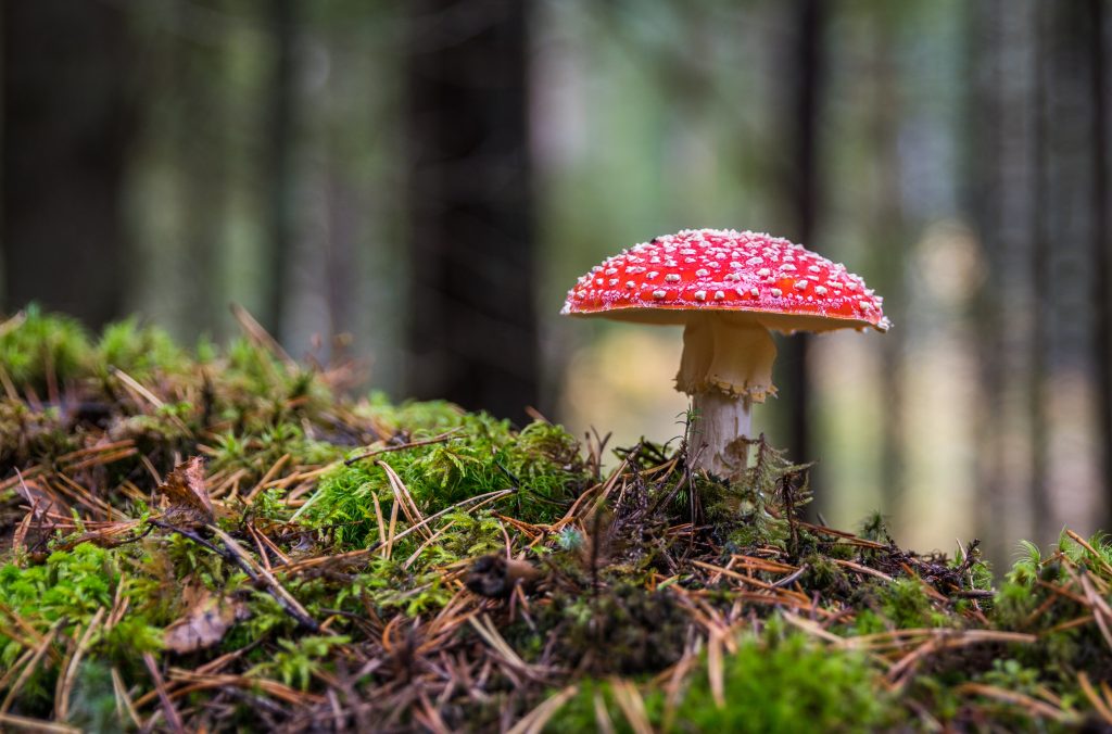
Saprotrophism
Together with bacteria, saprotrophic fungi are to a large extent responsible for the decomposition of organic matter. They are also responsible for the decay and decomposition of foodstuffs. Among other destructive saprotrophs are fungi that destroy timber and timber products as their mycelia invade and digest the wood; many of these fungi produce their spores in large, woody, fruiting bodies—e.g., bracket or shelf fungi. Paper, textiles, and leather are often attacked and destroyed by fungi. This is particularly true in tropical regions, where temperature and humidity are often very high.
The nutritional requirements of saprotrophs (and of some parasites that can be cultivated artificially) have been determined by growing fungi experimentally on various synthetic substances of known chemical composition. Fungi usually exhibit the same morphological characteristics in these culture media as they do in nature. Carbon is supplied in the form of sugars or starch; the majority of fungi thrive on such sugars as glucose, fructose, mannose, maltose, and, to a lesser extent, sucrose. Decomposition products of proteins, such as proteoses, peptones, and amino acids, can be used by most fungi as nitrogen sources; ammonium compounds and nitrates also serve as nutrients for many species. It is doubtful, however, that any fungus can combine, or fix, atmospheric nitrogen into usable compounds. Chemical elements such as phosphorus, sulfur, potassium, magnesium, and small quantities of iron, zinc, manganese, and copper are needed by most fungi for vigorous growth; elements such as calcium, molybdenum, and gallium are required by at least some species. Oxygen and hydrogen are absolute requirements; they are supplied in the form of water or are obtained from carbohydrates. Many fungi, deficient in thiamine and biotin, must obtain these vitamins from the environment; most fungi appear able to synthesize all other vitamins necessary for their growth and reproduction.
As a rule, fungi are aerobic organisms, meaning they require free oxygen in order to live. Fermentations, however, take place under anaerobic conditions. Knowledge of the physiology of saprotrophic fungi has enabled industry to use several species for fermentation purposes. One of the most important groups of strictly anaerobic fungi are members of the genera Neocallimastix (phylum Neocallimastigomycota), which form a crucial component of the microbial population of the rumen of herbivorous mammals. These fungi are able to degrade plant cell wall components, such as cellulose and xylans, that the animals cannot otherwise digest.
Parasitism in plants and insects
In contrast with the saprotrophic fungi, parasitic fungi attack living organisms, penetrate their outer defenses, invade them, and obtain nourishment from living cytoplasm, thereby causing disease and sometimes death of the host. Most pathogenic (disease-causing) fungi are parasites of plants. Most parasites enter the host through a natural opening, such as a stoma (microscopic air pore) in a leaf, a lenticel (small opening through bark) in a stem, a broken plant hair or a hair socket in a fruit, or a wound in the plant. Among the most common and widespread diseases of plants caused by fungi are the various downy mildews (e.g., of grape, onion, tobacco), the powdery mildews (e.g., of grape, cherry, apple, peach, rose, lilac), the smuts (e.g., of corn, wheat, onion), the rusts (e.g., of wheat, oats, beans, asparagus, snapdragon, hollyhock), apple scab, brown rot of stone fruits, and various leaf spots, blights, and wilts. These diseases cause great damage annually throughout the world, destroying many crops and other sources of food. For example, nearly all the chestnut forests of the United States have been destroyed by the chestnut blight fungus (Cryphonectria parasitica), and the elms in both the United States and Europe have been devastated by Ophiostoma ulmi, the fungus that causes Dutch elm disease.
Infection of a plant takes place when the spores of a pathogenic fungus fall on the leaves or the stem of a susceptible host and germinate, each spore producing a germ tube. The tube grows on the surface of the host until it finds an opening; then the tube enters the host, puts out branches between the cells of the host, and forms a mycelial network within the invaded tissue. The germ tubes of some fungi produce special pressing organs called appressoria, from which a microscopic, needlelike peg presses against and punctures the epidermis of the host; after penetration, a mycelium develops in the usual manner. Many parasitic fungi absorb food from the host cells through the hyphal walls appressed against the cell walls of the host’s internal tissues. Others produce haustoria (special absorbing structures) that branch off from the intercellular hyphae and penetrate the cells themselves. Haustoria, which may be short, bulbous protrusions or large branched systems filling the whole cell, are characteristically produced by obligate (i.e., invariably parasitic) parasites; some facultative (i.e., occasionally parasitic) parasites also produce them. Obligate parasites, which require living cytoplasm and have extremely specialized nutritional requirements, are exceptionally difficult, and often impossible, to grow in a culture dish in a laboratory. Examples of obligate parasites are the downy mildews, the powdery mildews, and the rusts.
Certain fungi form highly specialized parasitic relationships with insects. For example, the fungal genus Septobasidium is parasitic on scale insects (order Homoptera) that feed on trees. The mycelium forms elaborate structures over colonies of insects feeding on the bark. Each insect sinks its proboscis (tubular sucking organ) into the bark and remains there the rest of its life, sucking sap. The fungus sinks haustoria into the bodies of some of the insects and feeds on them without killing them. The parasitized insects are, however, rendered sterile. The perpetuation of the insect species and the spread of the fungus are accomplished by the uninfected members of the colony, which are protected from enemies by the fungus body. Newly hatched scale insects crawl over the surface of the fungus, which is at that time sporulating. Fungal spores adhere to the young insects and germinate. As the young insects settle down in a new place on the bark to begin feeding, they establish new fungal colonies. Thus, part of the insect colony is sacrificed to the fungus as food in return for the fungal protection provided for the rest of the insects. The insect is parasitic on the tree and the fungus is parasitic on the insect, but the tree is the ultimate victim.
The sooty molds constitute another interesting ecological group of fungi that are associated with insects. The majority of sooty molds are tropical or subtropical, but some species occur in the temperate zones. All sooty molds are epiphytic (i.e., they grow on the surfaces of other plants), but only in areas where scale insects are present. The fungi parasitize neither the plants nor the insects but rather obtain their nourishment exclusively from the honeydew secretions of the scale insects. Growth of the dark mycelium over the plant leaves, however, is often so dense as to significantly reduce the intensity of the light that reaches the leaf surface; this reduction in turn significantly reduces the rate of photosynthesis. Insect-fungus associations found in the tropical forests of Central and South America include the unique relationship of leafcutter ants (sometimes called parasol ants) with fungi in the family Lepiotaceae (phylum Basidiomycota). The ants cultivate the fungi in their nests as an ongoing food supply and secrete enzymes that stimulate or suppress the growth of the fungi.
Source
https://en.wikipedia.org/wiki/Fungus
https://www.britannica.com/science/fungus/Parasitism-in-plants-and-insects
https://www.usu.edu/herbarium/education/fun-facts-about-fungi/what-are-fungi
https://microbiologysociety.org/why-microbiology-matters/what-is-microbiology/fungi.html